Melanoma 101: Basics of Melanoma

When a doctor first informed me that I have superficial spreading malignant
melanoma, I was shocked. While the dermatologist was trying to reassure me that
the cancer was caught very early and not to worry, I was terrified! I was not even in high school yet and I was having a medical crisis. The fear
soon grew into frustration as I was refused to be told what melanoma was and
how the cancer/treatment would progress over the long term. In this blog I hope
to do what I wished my team of dermatologists would have done back then. With research and facts, I will explain what melanoma is, how it
progresses, what the risk factors are, and prevention. My goal is to assist
people recently diagnosed and their families to gain clarity.
Knowing the anatomy of skin is essential to understanding what melanoma is and how it
forms. Skin can be divided into two distinct layers, the epidermis and dermis.
The epidermis, which is the first layer of your skin, is made of rows and rows
of cells that are on top of each other. The rows of cells slowly become more
and more compact as they get farther away from the body. The cells eventually
start to die off once they reach the very top layer. In between these cells
that form the epidermis, are the cells that give the skin its color. These
cells are called melanocytes. Melanocytes
produce the pigmentation of our skin called melanin. While everyone has the
same number of melanocytes in their skin, the amount of melanin is different.
When the skin is exposed to UV light more melanin is produced giving a
darker/tan appearance. (Montagna) The second layer of skin is called the dermis. The dermis is richly supplied by blood vessels and a
web of lymph vessels. These two pipe systems are vital in cooling and cycling
nutrients throughout the body. Hair, lymph nodes, and sweat glands are all
found in the dermis. (Montagna) .

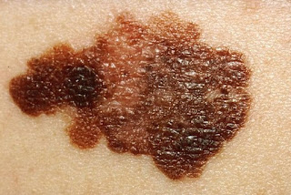
When the skin is exposed to UVA and UVB light, the melanocytes produce more pigmentation to protect the skin from becoming burned and damaged. The pigmentation that is produced usually results in benign moles and or freckles on the skin. But, when a melanocyte DNA becomes damaged from UVA and UVB, it can cause the cell to begin to replicate uncontrolled, produce more pigmentation then normal, and as a result forming melanoma. These cancer cells will then edge out other cells around it and spread deeper and wider in the skin tissue. The deeper the cancer gets to the dermis the more nutrients it has access to replicate more with. Once the cancer forms a tumor in the skin it can later spread to the lymph nodes. Once cancer is present in the lymph nodes, it can then spread to the rest of the body.
Works Cited
Montagna, F. John G. Ebling and William. 1.
Eleven April 2016. Eleven Febuary 2018.
Rastrelli, Marco. "Melanoma M (Zero): Diagnosis
and Therapy." ISRN Dermatology (2013): 10. Document.

Comments
Post a Comment